

Review - DOI:10.33594/000000742
Accepted 30 August, 2023 - Published online : 12 November, 2024
1Institute of Neurointervention/ 2Department of Neurology, Christian Doppler University Hospital, Paracelsus Medical University, Salzburg Austria,
3Department of Neuroradiology, Medical University of Innsbruck, 6020 Innsbruck, Austria,
4Department of Diagnostic and Interventional Neuroradiology, University Medical Center Hamburg-Eppendorf, Germany
Background/Aims: Intrasaccular devices are an emerging treatment for cerebral aneurysms. However, current grading scales for outcome assessment are difficult to apply as device positioning is not taken into account. We present a novel grading scale to assess how likely a complete occlusion is predictale for cerebral aneurysms treated with intrasaccular devices. Materials: The scale was developed using results from 143 aneurysms treated at our institution with intrasaccular devices from 2019 to 2023. Angiographic images and clinical complications were taken to illustrate key aspects of the scale. Results: The scale considers device position relative to the parent artery and the aneurysm wall, contrast filling, neck coverage, and contrast inflow/stability. Conclusions: This scale helps standardize outcome measurements in accordance with the Modified Raymond–Roy Classification and O`Kelly Marotta grading scales, providing a basis for the common reporting of results.
Intrasaccular devices are new and promising tools for treating wide-neck and bifurcation intracranial aneurysms.1-4 Currently, no aneurysm grading scale is compatible with the intrasaccular devices such as the Contour device (Cerus Endovascular, Fremont, California, USA) and the Artisse (Medtronic, Irvine, California, USA). The Raymond–Roy Occlusion Classification and O’Kelly-Marotta grading scales are difficult to apply to aneurysms treated with intrasaccular devices that differ in position and function.5, 6
Only two grading scales exist to score treatment with intrasaccular devices, both used in the context of the Woven EndoBridge device: the Woven EndoBridge Occlusion Scale and the Bicêtre Occlusion Scale Score.7, 8 Both scales grade occlusion based on contrast filling and stasis inside the aneurysm sac and indicate the device position to the aneurysm wall;7-9 however, these scales fail to measure key variables that can indicate the likelihood of successful occlusion and/or the need for re-treatment or medication.7, 8 To successfully grade treatment with intrasaccular devices, a scale should indicate device stability inside the aneurysm, device position at the aneurysm wall and neck, and the presence of device protrusion into the parent artery. Here, we propose a novel grading scale for aneurysms treated with intrasaccular devices applicable to both bifurcation and sidewall aneurysms.
The scale was developed using results from our animal experience with WEB, Contour and Artisse ( n = 50 aneurysms) and consecutive patients (n = 93), treated with these intrasaccular devices for cerebral aneurysms at our institution from June 2019 to June 2023. In some cases, adjunctive treatments such as stent placement or coiling were necessary, based on the experience of thromboembolic complications, parent artery occlusions due to bulging of the device into the parent artery, or recurrences during followup periods. Angiographic images were obtained and evaluated using digital subtraction angiography (DSA). For follow-up imaging coan beam CT and DSA was used. A retrospective analysis of these data including longterm followup was used to devellop the grading scale.
Angiographic images were obtained and evaluated using DSA. Cone beam CT and DSA were used for follow-up imaging.
The new grading scale uses one of four letter grades (A-D) to indicate the amount of contrast filling, combined with a number grade (1-4) to indicate device position. Additionally, the score distinguishes between different device positions in relation to the parent artery.
Contrast Filling Grade
Contrast filling of the aneurysm sac is graded using a four-letter scale similar to the O’Kelly-Marotta Scale: A indicates complete filling (>95%); B indicates incomplete filling (5-95%); C indicates the presence of a neck remnant (<5%); and D indicates no filling (0%). Fig. 1 shows a schematic representation of these four grades in the aneurysm and in the aneurysm in relation to the parent artery.
Device position
Device position is indicated using a numerical grade of 1-3, similar to Raymond-Roy Occlusion Classification. Grade 1 indicates perfect neck reconstruction with only the proximal marker protruding into the target artery, while Grade 2 indicates a neck remnant - device with proximal marker is inside the sac. Grade 3 indicates inflow into the aneurysm sac, a dog leg, or that the device position is not stable, indicating an unacceptable device position – the selected device was probably too small or too large. Fig. 2 shows a schematic representation of the device position within sidewall aneurysms for each of the three grades. Fig. 3 shows a schematic representation of the device position within bifurcation aneurysms for each of the three grades.
Table 1 provides an interpretation of all scale grades, possible prognosis, and medication strategy sugesstions for each in accordance to our experience. The grade can indicate whether aneurysm occlusion is likely, and whether antiplatelet treatment might become necessary.Illustrative angiographic examples of patients treated with Contour devices are shown in Figures 4 and 5.
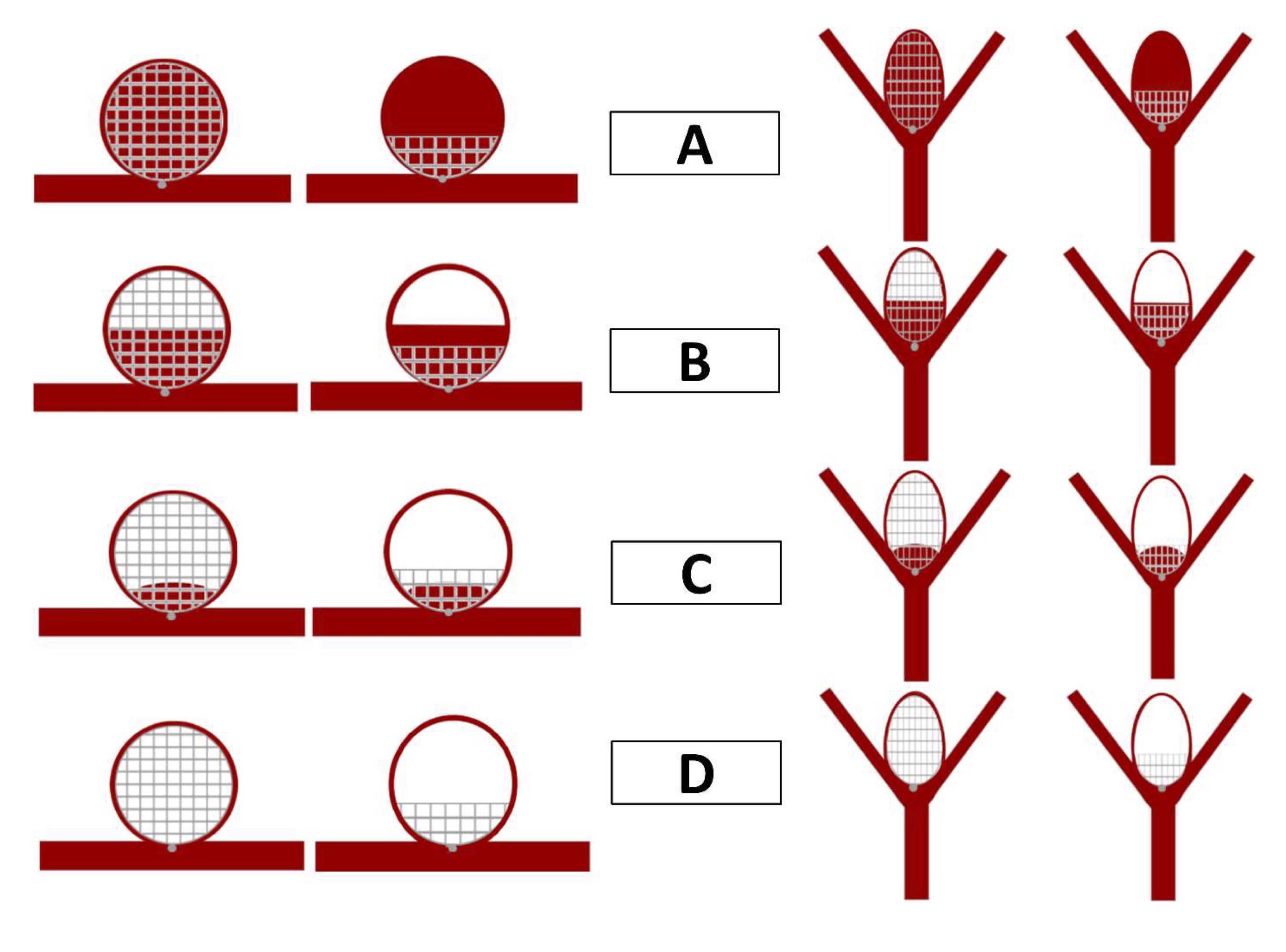
Fig. 1. Contrast Filling. As with the O’Kelly-Marotta scale, the contrast filling of the aneurysm sac is graded: A, complete (>95%); B, incomplete (5-95%); C, neck remnant (<5%); or D, no filling (0%). The left sided drawings show the devices in sidewall aneurysms and the right sided drawings in bifurcation aneurysms.
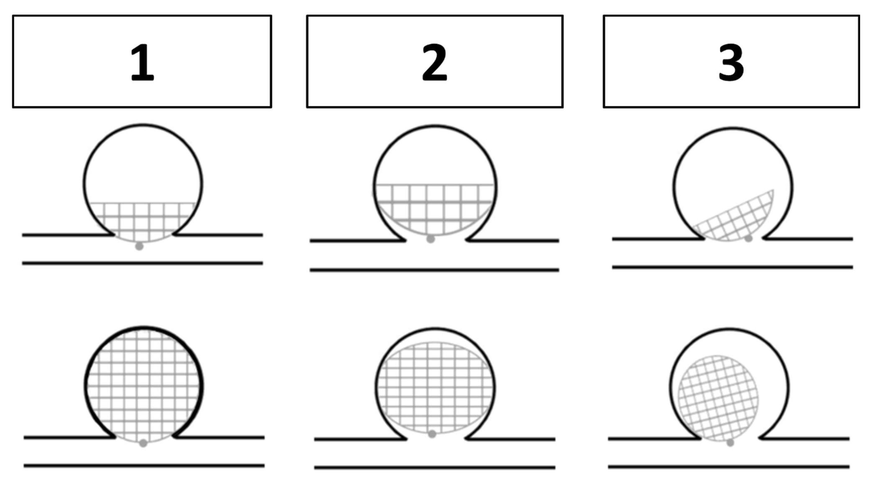
Fig. 2. Device Position in Relation to Parent Artery within Sidewall Aneurysms. Grades (Grade 1-3) indicating neck coverage and device stability in relation to the parent artery. Grade 1, perfect neck reconstruction with only the proximal marker protruding into the target artery – including a slight protrusion of the device into the parent artery; Grade 2, neck remnant; Grade 3, any inflow into the aneurysm sac outside of the device, a dog leg or a protrusion into the parent artery with narrowing of the artery lumen, all indicating that the device position is not stable or can cause thromboembolic complications.
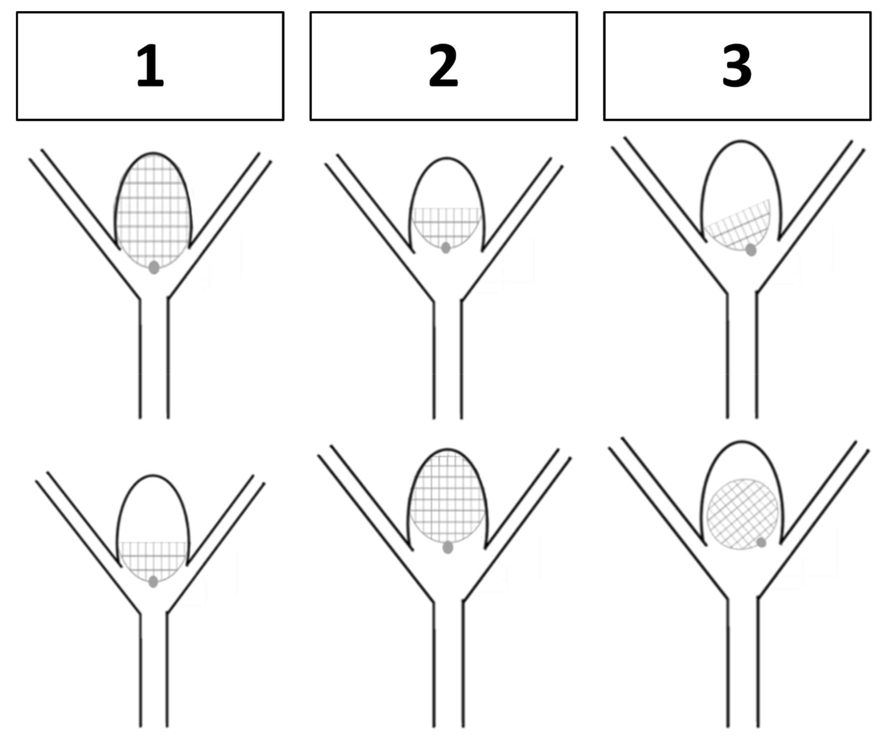
Fig. 3. Device Position in Relation to Parent Artery within Bifurcation Aneurysms. Grades (Grade 1-3) indicating neck coverage and device stability in relation to the parent artery. Grade 1, perfect neck reconstruction with only the proximal marker protruding into the target artery – including a slight protrusion of the device into the parent artery; Grade 2, neck remnant; Grade 3, any inflow into the aneurysm sac outside of the device, a dog leg or a protrusion into the parent artery with narrowing of the artery lumen, all indicating that the device position is not stable or can cause thromboembolic complications.
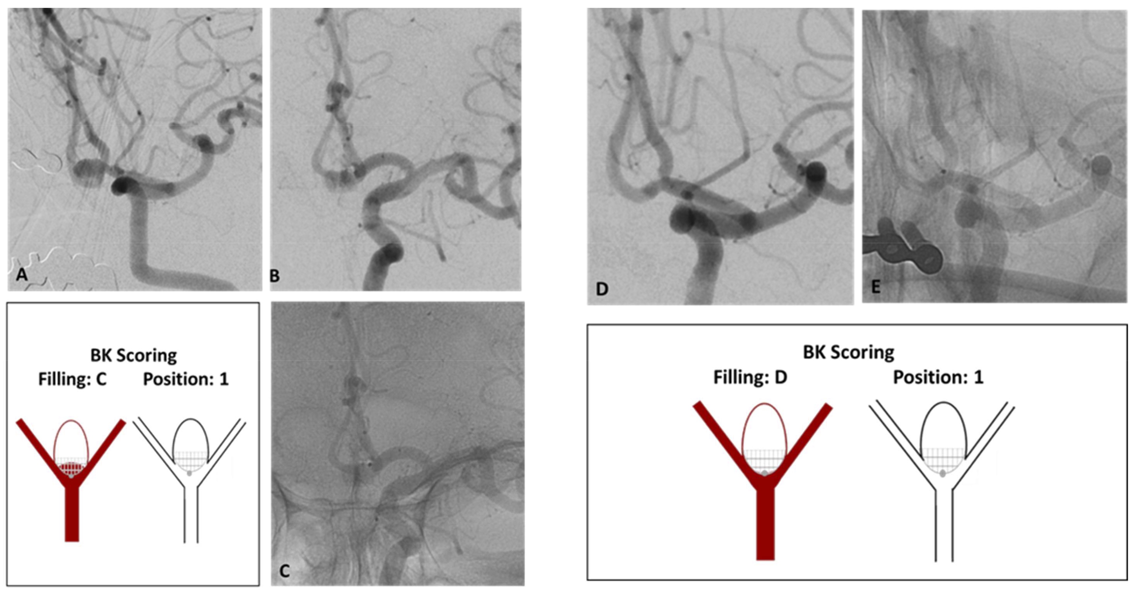
Fig. 4. 72-year-old male with incidental anterior communicating artery (Acom) aneurysm. A) The untreated Acom aneurysm measuring 5 x 6 mm. B-C) The aneurysm after treatment with Contour 7 mm with significant contrast stasis after detachment. Already, the aneurysm is two thirds smaller, and the image shows correct proximal marker position at the neck. D-E) At 6 months control the aneurysm is completely occluded with unchanged device position. Given the excellent device placement at the neck, occlusion was expected.
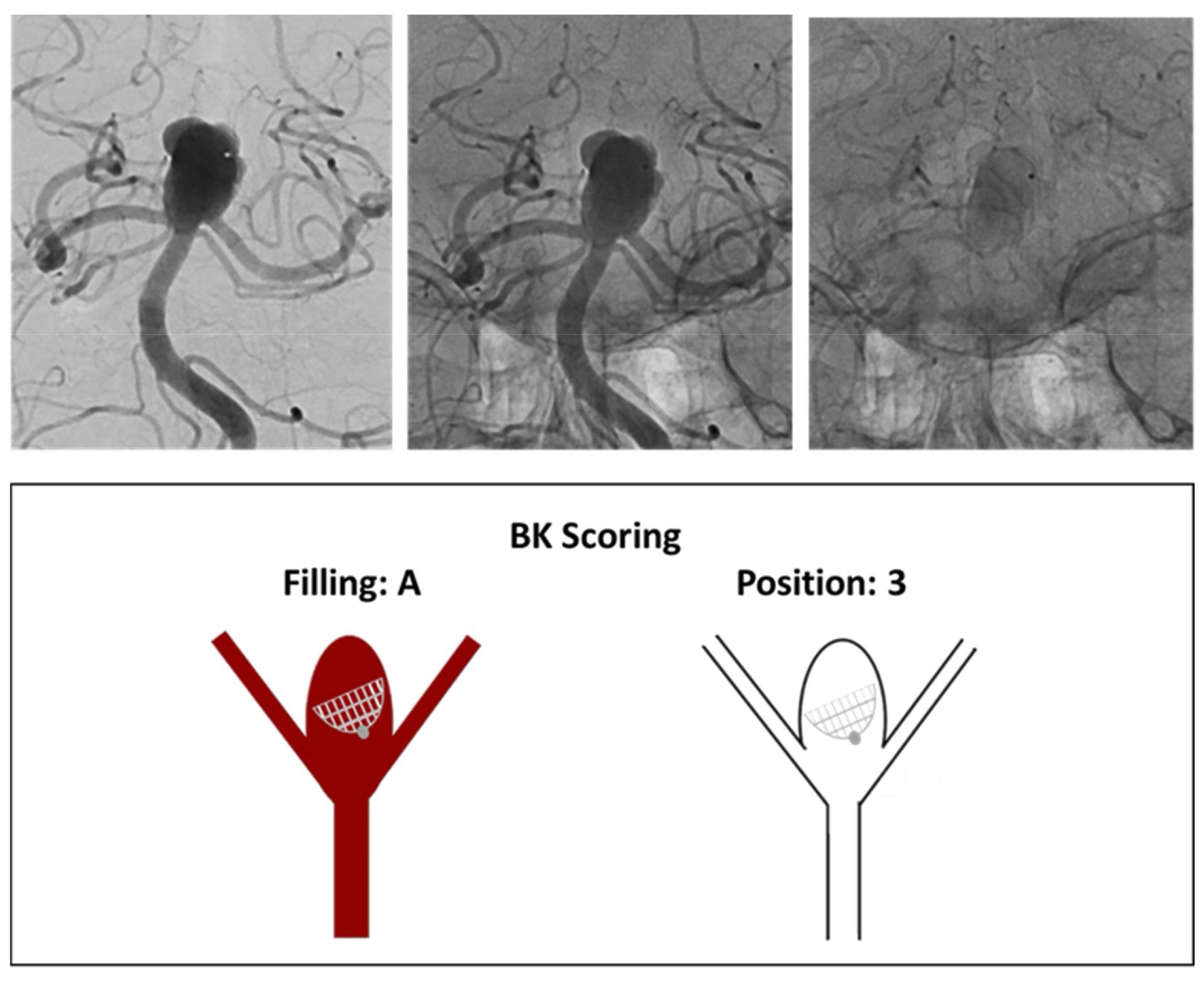
Fig. 5. 67-year-old patient, incidental finding of a basilar tip aneurysm. A) Basilar tip aneurysm measuring 9.0 x 10.0 mm treated with Contour 14. B) At one month control, device turned over resulting in an improper position during contrast filling inside the aneurysm sac. Due to the incorrect device position, the treatment is likely insufficient and may need to be repeated. IDOL Intrasaccular Device Occlusion and Location Scale; BOSS, Bicêtre Occlusion Scale Score; MRRC, modified Raymond–Roy Classification; RROC, Raymond–Roy Occlusion Classification; WOS, WEB Occlusion Scale
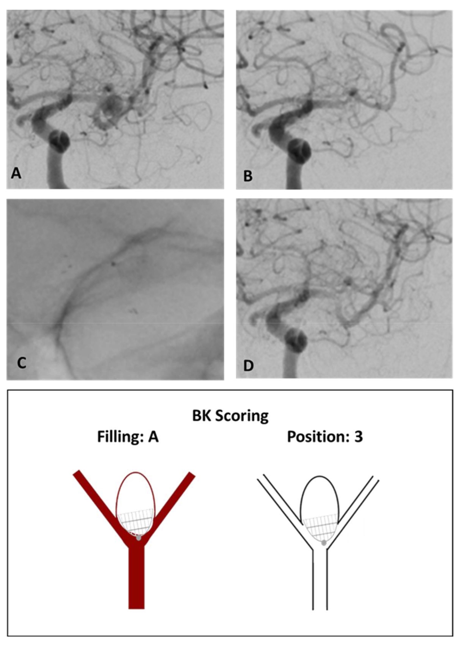
Fig. 6. 68-year-old female with incidental finding of an a middle cerebral arterial (MCA) aneurysm, measuring 6.5 x 6.0 mm (A), treated with Contour 9 mm. After Contour deployment the inferior branch showed, beside immediate aneurysm occlusion, a significant reduced flow (B), because of Contour system comprising the inferior branch, so that additionally a Neuroform Stent (3.5x15) was implanted to keep the branch open (C). After stent implantation the branch was open and the aneurysm remained occluded (D).
We present a novel grading scale for sidewall and bifurcation aneurysms treated with intrasaccular devices. The scale is designed to standardize aneurysm occlusion grading and intrasaccular device position in relation to the parent artery, which is critical in determining the need for re-treatment or changes in medication.
Current grading scales for intrasaccular devices do not adequately consider device position.7-9 If an aneurysm continues to show complete filling of the aneurysm sac after treatment with an intrasaccular device, but otherwise shows excellent device position at the neck, it would receive a score of “1 A” with our scale, indicating that the aneurysm is well-treated with occlusion predicted over the follow-up periods. This same aneurysm would receive a score of III using the Raymond-Roy Occlusion Classification, and either IIIa or IIIb using the modified Raymond–Roy classification;6, 10 both indicating insufficient embolization. The only Woven EndoBridge Occlusion Scale grade that would be appropriate is D, indicating contrast opacification extending beyond the aneurysm neck.7 The Bicêtre Occlusion Scale Score (3 or 3+1) would also indicate poor results.8 Therefore, all current grading scales would suggest disappointing results, while the aneurysm is likely well-treated and will presumably occlude over time.
Treatment using intrasaccular devices is likely to increase, underscoring the need for an appropriate and consistent grading system.
We present a novel scale that characterizes results after aneurysm treatment that incorporates the position of the device in an attempt to help standardize outcome measurements in accordance with the currently used grading scales.
The authors acknowledge Superior Medical Experts for editorial assistance.
Author contributions
MKO: Conception of the workEMTB: Data collection and drafting of the articleAG: Critical revision of the articleJF: Critical revision of the articleAll authors: validated the results independently and approved the final version of the manuscript.
Ethics approval for the animal studies and patient consent for each treatment are available. No ethical approval as a retrospective data analysis.
Funding
No external funding was utilized for this study or manuscript preparation.
No competing interests related to the methods or materials used in this study.